several spectroscopic methodologies. Among these, high-resolution 1H NMR spectroscopy coupled with
pattern recognition is a method of great success due to its ability to quantify a large range of metabolites
simultaneously without preconceived ideas of the biomarkers associated with pathology. The use of
multivariate techniques such as PLS-Discriminant Analysis on a complex data provides a statistical tool
for discriminating between spectra from different classes of samples, thus reducing the large numbers of
spectral features to key metabolic perturbations.
In this study, 37 cerebrospinal fluid (CSF) samples (25 from patients with multiple sclerosis [MS]_and
12 from disease controls - idiopathic polyneuropathy and meningitis) were examined by 1H NMR
spectroscopy and the data analyzed by multivariate statistics. The study was approved by our local Ethics
Committee and all individuals gave informed consent. All CSF samples were collected for clinical
diagnostic purposes, and a small portion of the sample was kept for 1H-NMR analyses. All 1H NMR
spectra were acquired at 499.9 MHz using a INOVA 500 spectrometer (Varian) and a 5 mm triple
resonance inverse probe. Spectra were recorded at 298 K and represented the sum of 64 transient
acquired over64 K data points with a spectral width of 10 kHz. FID were transformed using 1 degree of
zero filling and 0.5 Hz exponential multiplication. The reference was 2.5 mM (TPS) at m=0.0 was added
(aqueous solution 100 ml, 2.5 mM) to the CSF sample (500 mL). All spectra were treated prior to the
multivariate statistics and pattern recognition by adjusting the TPS peak for possible shift and to the same
height. Each speactum was basline corrected using a linear fit and the final data set was autoscaled. The
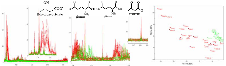
1H NMR spectra demonstrated resonance arising from acetate, alanine, b-hydroxybutyrate, citrate,
formate, glucose, glutamine, glutamate, myo-inositol, isobutyrate, lactate, succinate, tyrosine and valine.
PLS-DA demonstrated that CSF from MS and disease control patients were different with increased,
glutamine, glutamate, b-hydroxybutirate and acetoacetate in patients with MS (Figure 1a and b). Scores
plot (Figure 1c) discriminates the group with MS from the disease control group. Leave-one-out
crossvalidation indicated only one misclassification.
The increase in glutamine might be related to aminoacids degradation, while b-hydroxybutirate and
acetylacetate are ketonic bodies which are an alternative energetic route when glucose availability is low
or inefficient.